
What is a coronary angiogram?
A coronary angiogram is a diagnostic procedure that uses X-rays to see your heart’s blood vessels. Coronary angiograms are also called heart catheterization because of the long tube (catheter) that is used to see the coronary arteries. The doctors doing the angiogram can both diagnose and treat heart and blood vessel medical conditions on the same visit.
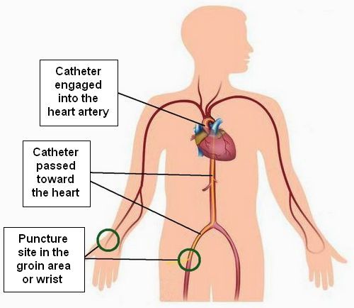
To perform an angiogram doctors first make a tiny hole in a blood vessel leading to the heart. This is usually done in the groin or wrist area. It allows the doctor to pass a long tube (catheter) all the way to the heart. After that*, X-ray contrast (also called ‘dye’) is injected into the blood vessels (arteries) of your heart. The X-ray machine shows the contrast and rapidly takes a series of pictures and videos. These pictures and videos allow the doctor to visualize what is going on inside your blood vessels. Depending on what is seen, they can perform procedures to open arteries that are partially or completely blocked.
How does an angiogram look like?
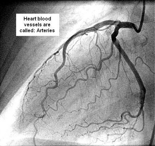
The angiogram is obtained in black and white and looks like this:
Heart Anatomy
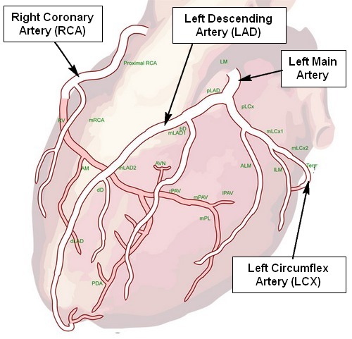
There are three main coronary arteries that supply the heart muscle. The Right Coronary Artery (RCA) that supplies blood to the right side and the bottom of the heart. The Left Main artery has two major branches called the Left Anterior Descending (LAD) Artery and the Left Circumflex (LCx) Artery. These two branches supply the front and the left side of the heart. We use the diagram below to describe what is seen in your angiogram. A copy of this diagram is given to our patients at the end of their procedure.
Read next: